Fichier:Wuchereria bancrofti LifeCycle.gif

Le contenu de la page n’est pas pris en charge dans d’autres langues.
Une page de Wikipédia, l'encyclopédie libre.

Wuchereria_bancrofti_LifeCycle.gif(513 × 435 pixels, taille du fichier : 33 kio, type MIME : image/gif)

Ce fichier et sa description proviennent de Wikimedia Commons.

Description

Wuchereria bancrofti

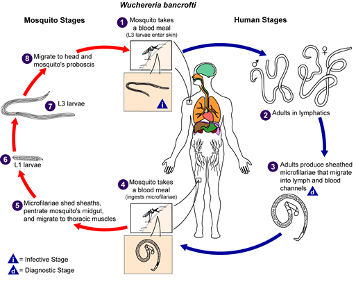
Life cycle of Wuchereria bancrofti

Different species of the following genera of mosquitoes are vectors of W. bancrofti filariasis depending on geographical distribution. Among them are: Culex (C. annulirostris, C. bitaeniorhynchus, C. quinquefasciatus, and C. pipiens); Anopheles (A. arabinensis, A. bancroftii, A. farauti, A. funestus, A. gambiae, A. koliensis, A. melas, A. merus, A. punctulatus and A. wellcomei); Aedes (A. aegypti, A. aquasalis, A. bellator, A. cooki, A. darlingi, A. kochi, A. polynesiensis, A. pseudoscutellaris, A. rotumae, A. scapularis, and A. vigilax); Mansonia (M. pseudotitillans, M. uniformis); Coquillettidia (C. juxtamansonia). During a blood meal, an infected mosquito introduces third-stage filarial larvae onto the skin of the human host, where they penetrate into the bite wound. They develop in adults that commonly reside in the lymphatics. The female worms measure 80 to 100 mm in length and 0.24 to 0.30 mm in diameter, while the males measure about 40 mm by .1 mm. Adults produce microfilariae measuring 244 to 296 μm by 7.5 to 10 μm, which are sheathed and have nocturnal periodicity, except the South Pacific microfilariae which have the absence of marked periodicity. The microfilariae migrate into lymph and blood channels moving actively through lymph and blood. A mosquito ingests the microfilariae during a blood meal. After ingestion, the microfilariae lose their sheaths and some of them work their way through the wall of the proventriculus and cardiac portion of the mosquito's midgut and reach the thoracic muscles. There the microfilariae develop into first-stage larvae and subsequently into third-stage infective larvae. The third-stage infective larvae migrate through the hemocoel to the mosquito's prosbocis and can infect another human when the mosquito takes a blood meal.
Source http://www.dpd.cdc.gov/dpdx/images/ParasiteImages/A-F/Filariasis/W_bancrofti_LifeCycle.gif
Auteur
Autorisation
(Réutilisation de ce fichier)
Public domain
Cette image est l’œuvre des
Centers for Disease Control and Prevention
, division du Département de la Santé et des Services Sociaux des États-Unis, réalisée par un employé dans le cadre de ses activités professionnelles. En tant qu'œuvre du gouvernement fédéral des États-Unis d'Amérique, cette image est placée dans le domaine public.

eesti  Deutsch  čeština  español  português  English  français  Nederlands  polski  slovenščina  suomi  македонски  українська  日本語  中文(简体)‎  中文(繁體)‎  العربية  +/−

Légendes

Ajoutez en une ligne la description de ce que représente ce fichier

Éléments décrits dans ce fichier

dépeint

image/gif

Historique du fichier

Cliquer sur une date et heure pour voir le fichier tel qu'il était à ce moment-là.

Date et heureVignetteDimensionsUtilisateurCommentaire
actuel5 novembre 2008 à 16:15Vignette pour la version du 5 novembre 2008 à 16:15513 × 435 (33 kio)LycaonWatermark removed
14 mai 2006 à 23:02Vignette pour la version du 14 mai 2006 à 23:02513 × 435 (36 kio)Patho{{Information| |Description=Filariasis [Brugia malayi] [Brugia timori] [Loa loa] [Mansonella ozzardi] [Mansonella perstans] [Mansonella streptocerca] [Onchocerca volvulus] [Wuchereria bancrofti] Life cycle of Wuchereria bancrofti Different species

Les 3 pages suivantes utilisent ce fichier :

Usage global du fichier

Les autres wikis suivants utilisent ce fichier :