
Fichier:Long bone assigned to Pterodactylus cuvieri.jpg
Fichier d’origine (4 740 × 1 428 pixels, taille du fichier : 4,46 Mio, type MIME : image/jpeg)
Ce fichier et sa description proviennent de Wikimedia Commons.
Description
| Description |
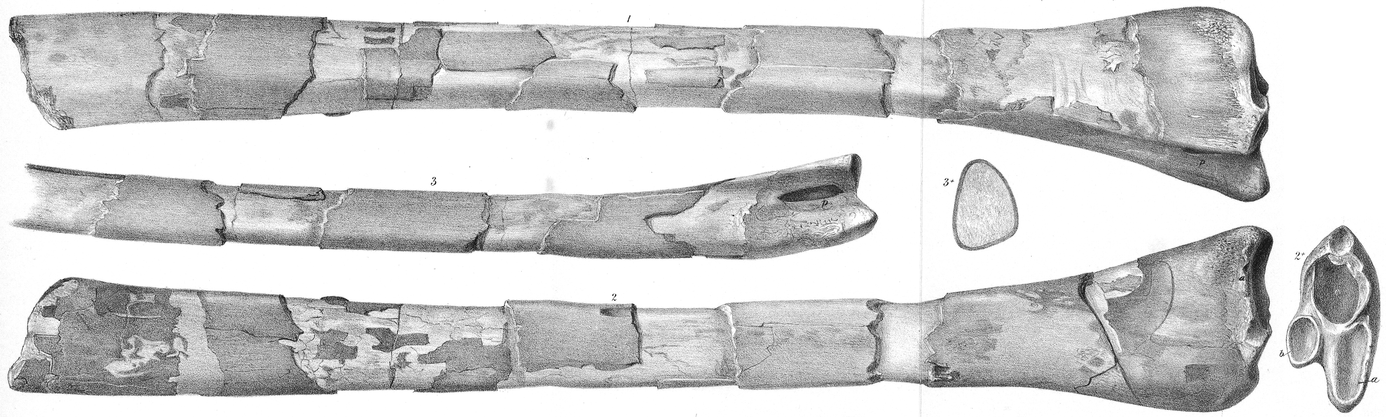
Figs. 1 and 2. Wing-phalanx bone of Pterodactylus Cuvieri. Fig. 2*. Articular end of ditto: a and b, articular surfaces; c, fractured surface leading to the cavity of the bone. Fig. 3. Portion of the narrowest side of the same bone, showing the pneumatic foramen at p. Fig. 3*. Section of the same bone four inches from the articular end, showing the thickness of its dense osseous wall, and the wide air-cavity. From the Burham Chalk-pit, Kent. In the Collection of J. Toulmin Smith, Esq. |
| Date | entre 1849 et 1884 |
| Source | https://www.biodiversitylibrary.org/item/99821#page/25/mode/1up |
| Auteur | Joseph Dinkel |
Conditions d’utilisation
|
Cette œuvre est également dans le domaine public dans tous les pays pour lesquels le droit d’auteur a une durée de vie de 70 ans ou moins après la mort de l’auteur. Cette œuvre est dans le domaine public aux États-Unis car elle a été publiée avant le 1er janvier 1929. | |
| Ce fichier a été identifié comme étant exempt de restrictions connues liées au droit d’auteur, y compris tous les droits connexes et voisins. | |
Historique du fichier
Cliquer sur une date et heure pour voir le fichier tel qu'il était à ce moment-là.
| Date et heure | Vignette | Dimensions | Utilisateur | Commentaire | |
|---|---|---|---|---|---|
| actuel | 16 février 2021 à 23:43 | 4 740 × 1 428 (4,46 Mio) | FunkMonk | {{Information |Description=Figs. 1 and 2. Wing-bone of Pterodactylus Cuvieri. Fig. 2*. Articular end of ditto: a and b, articular surfaces; c, fractured surface leading to the cavity of the bone. Fig. 3. Portion of the narrowest side of the same bone, showing the pneumatic foramen at p. Fig. 3*. Section of the same bone four inches from the articular end, showing the thickness of its dense osseous wall, and the wide air-cavity. From the Burham Chalk-pit, Kent. In the Collection of J. Toulm... |
Utilisation du fichier
La page suivante utilise ce fichier :
Usage global du fichier
Les autres wikis suivants utilisent ce fichier :
- Utilisation sur en.wikipedia.org