
Fichier:Echinococcus Life Cycle.png
Echinococcus_Life_Cycle.png (600 × 571 pixels, taille du fichier : 44 kio, type MIME : image/png)
Ce fichier et sa description proviennent de Wikimedia Commons.
Description
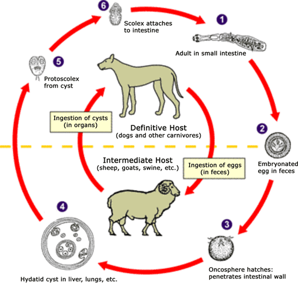
The adult Echinococcus granulosus (3 to 6 mm long) [1] resides in the small bowel of the definitive hosts (dogs or other carnivores). Gravid proglottids release eggs [2] that are passed in the feces. After ingestion by a suitable intermediate host (under natural conditions: sheep, goat, swine, cattle, horses, camel), the egg hatches in the small bowel and releases an oncosphere [3] that penetrates the intestinal wall and migrates through the circulatory system into various organs, especially the liver and lungs. In these organs, the oncosphere develops into a cyst [4] that enlarges gradually, producing protoscolices and daughter cysts that fill the cyst interior. The definitive host becomes infected by ingesting the cyst-containing organs of the infected intermediate host. After ingestion, the protoscolices [5] evaginate, attach to the intestinal mucosa [6] and develop into adult stages [1] in 32 to 80 days. The same life cycle occurs with E. multilocularis (1.2 to 3.7 mm), with the following differences: the definitive hosts are foxes, and to a lesser extent dogs, cats, coyotes and wolves; the intermediate host are small rodents; and larval growth (in the liver) remains indefinitely in the proliferative stage, resulting in invasion of the surrounding tissues. With E. vogeli (up to 5.6 mm long), the definitive hosts are bush dogs and dogs; the intermediate hosts are rodents; and the larval stage (in the liver, lungs and other organs) develops both externally and internally, resulting in multiple vesicles. E. oligarthrus (up to 2.9 mm long) has a life cycle that involves wild felids as definitive hosts and rodents as intermediate hosts. Humans become infected by ingesting eggs , with resulting release of oncospheres in the intestine and the development of cysts in various organs.
Image adapted from original available at the United States Centres for Disease Control Parasitology Identification Laboratory ([1] copie d'archive at the Wayback Machine).

|
Une version vectorielle de cette image existe, dans le format « SVG ». Si elle n’est pas inférieure, elle devrait être utilisée à la place de la présente version pour des affichages en plus grandes dimensions ou nécessitant une meilleure résolution.
File:Echinococcus Life Cycle.png → File:Echinococcus Life Cycle.svg
Pour plus d’informations sur les images vectorielles, consultez la page de transition de Commons vers le format SVG. Voir aussi les informations à propos de la manière dont le logiciel MediaWiki gère les images au format SVG. |
 |
Conditions d’utilisation
Cette image est l’œuvre des
Centers for Disease Control and Prevention , division du Département de la Santé et des Services Sociaux des États-Unis, réalisée par un employé dans le cadre de ses activités professionnelles. En tant qu'œuvre du gouvernement fédéral des États-Unis d'Amérique, cette image est placée dans le domaine public.
|
Historique du fichier
Cliquer sur une date et heure pour voir le fichier tel qu'il était à ce moment-là.
| Date et heure | Vignette | Dimensions | Utilisateur | Commentaire | |
|---|---|---|---|---|---|
| actuel | 24 janvier 2007 à 12:54 | | 600 × 571 (44 kio) | Pngbot | optimized with optipng |
| 26 avril 2005 à 03:48 |  | 600 × 571 (55 kio) | FirstPrinciples~commonswiki | Smaller & clearer | |
| 26 avril 2005 à 03:36 |  | 800 × 761 (79 kio) | FirstPrinciples~commonswiki |
Utilisation du fichier
Aucune page n’utilise ce fichier.