
Fichier:Nematocyst discharge.png
Pas de plus haute résolution disponible.
Nematocyst_discharge.png (480 × 371 pixels, taille du fichier : 190 kio, type MIME : image/png)
Ce fichier et sa description proviennent de Wikimedia Commons.

|
Cette image (de type biologie) devrait être recréée dans un format vectoriel, en tant que fichier SVG. Cela offrirait plusieurs avantages : voir Commons:Media for cleanup pour plus d'informations. Si une version SVG de cette image est déjà disponible, merci de bien vouloir l'envoyer. Après cela, remplacez ce modèle par {{vector version available|nouveau nom d'image.svg}}.
|
Description
| Description |
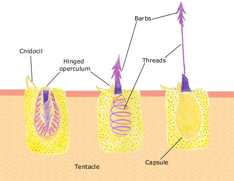
English: The diagram above shows the anatomy of a nematocyst cell and its “firing” sequence, from left to right. On the far left is a nematocyst inside its cellular capsule. The cell’s thread is coiled under pressure and wrapped around a stinging barb. When potential prey makes contact with the tentacles of a polyp, the nematocyst cell is stimulated. This causes a flap of tissue covering the nematocyst—the operculum—to fly open. The middle image shows the open operculum, the rapidly uncoiling thread and the emerging barb. On the far right is the fully extended cell. The barbs at the end of the nematocyst are designed to stick into the polyp’s victim and inject a poisonous liquid. When subdued, the polyp’s tentacles move the prey toward its mouth and the nematocysts recoil back into their capsules. |
| Date | 11 avril 2007 (date de téléversement originale) |
| Source | Transféré de en.wikipedia à Commons. |
| Auteur | Original téléversé par Spaully sur Wikipédia anglais. |
Conditions d’utilisation
Ce fichier est disponible selon les termes de la licence Creative Commons Partage des Conditions Initiales à l'Identique 1.0 Générique.
Creative Commons a retiré cet outil juridique et recommande de ne pas l’appliquer aux œuvres.
|
Cette image est dans le domaine public car son contenu provient de la
National Oceanic and Atmospheric Administration , réalisé par un employé dans le cadre de ses activités professionnelles.
|
Journal des téléversements d’origine
La page de description originale était ici. Tous les noms d'utilisateur qui suivent se rapportent à en.wikipedia.
- 2007-04-11 17:10 Spaully 480×371×8 (194868 bytes) Modified from: http://www.oceanservice.noaa.gov/education/kits/corals/media/supp_coral01b.html {{Information |Description=Nematocyst discharge process. |Source= Modified from [http://www.oceanservice.noaa.gov/education/kits/corals/media/supp_coral01b.html
Historique du fichier
Cliquer sur une date et heure pour voir le fichier tel qu'il était à ce moment-là.
| Date et heure | Vignette | Dimensions | Utilisateur | Commentaire | |
|---|---|---|---|---|---|
| actuel | 13 octobre 2007 à 19:29 | | 480 × 371 (190 kio) | Alison | {{Information |Description===Description== The diagram above shows the anatomy of a nematocyst cell and its “firing” sequence, from left to right. On the far left is a nematocyst inside its cellular capsule. The cell’s thread is coiled under pressur |
Utilisation du fichier
La page suivante utilise ce fichier :
Usage global du fichier
Les autres wikis suivants utilisent ce fichier :
- Utilisation sur ca.wikipedia.org
- Utilisation sur ceb.wikipedia.org
- Utilisation sur en.wikipedia.org
- Utilisation sur hr.wikipedia.org
- Utilisation sur id.wikipedia.org
- Utilisation sur it.wikibooks.org
- Utilisation sur ja.wikipedia.org
- Utilisation sur lv.wikipedia.org
- Utilisation sur ms.wikipedia.org
- Utilisation sur my.wikipedia.org
- Utilisation sur pa.wikipedia.org
- Utilisation sur pt.wikipedia.org
- Utilisation sur simple.wikipedia.org
- Utilisation sur sv.wikipedia.org
- Utilisation sur te.wikipedia.org
- Utilisation sur th.wikipedia.org
- Utilisation sur vi.wikipedia.org