
Fichier:Bundleofhis.png
Bundleofhis.png (400 × 483 pixels, taille du fichier : 69 kio, type MIME : image/png)
Ce fichier et sa description proviennent de Wikimedia Commons.
Description
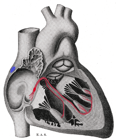
Schematic representation of the atrioventricular bundle of His. The bundle, represented in red, originates near the orifice of the coronary sinus, undergoes slight enlargement to form the AV node. The AV node tapers down into the bundle of HIS, which passes into the ventricular septum and divides into two bundle branches, the left and right bundles. Sometimes the 'left and right bundles of His' are called Purkyne or Purkinge fibres. The ultimate distribution cannot be completely shown in this diagram.
This image is misleading. Although it correctly places the SA and AV nodes in the right atrium, it appears as though there are only two papillary muscles in the right ventricle and three in the left ventricle. The opposite is actually true. The papillary muscles attach to chordae tendinae which then attach to the leaflets of the AV valves, preventing prolapse. The left AV valve is the mitral or bicuspid and only has two leaflets and therefore two papillary muscles. The right AV valve is the tricuspid and should have three papillary muscles corresponding to the three leaflets of the valve.
Conditions d’utilisation
Ce document est dans le domaine public aux États-Unis. Ceci s'applique aux travaux des États-Unis où le copyright a expiré, souvent parce que sa première publication s'est produite avant le 1er janvier 1929. Voir cette page pour davantage d'explication.
|
 | |
 |
Cette image peut ne pas être dans le domaine public en dehors des États-Unis (ceci s'applique particulièrement au Canada, en Chine (pas Hong Kong, Macao, ni Taïwan), en Allemagne ou en Suisse). Le créateur et l'année de la publication sont l'information essentielle et doivent être fournis.
|
Historique du fichier
Cliquer sur une date et heure pour voir le fichier tel qu'il était à ce moment-là.
| Date et heure | Vignette | Dimensions | Utilisateur | Commentaire | |
|---|---|---|---|---|---|
| actuel | 20 septembre 2006 à 19:40 | | 400 × 483 (69 kio) | Kauczuk | Bundle of His, from Gray's Anatomy 1918 |
Utilisation du fichier
La page suivante utilise ce fichier :
Usage global du fichier
Les autres wikis suivants utilisent ce fichier :
- Utilisation sur ar.wikipedia.org
- Utilisation sur az.wikipedia.org
- Utilisation sur bn.wikibooks.org
- Utilisation sur bs.wikipedia.org
- Utilisation sur ca.wikipedia.org
- Utilisation sur cs.wikipedia.org
- Utilisation sur de.wikipedia.org
- Utilisation sur de.wikibooks.org
- Utilisation sur el.wikipedia.org
- Utilisation sur en.wikipedia.org
- Utilisation sur en.wikibooks.org
- Utilisation sur es.wikipedia.org
- Utilisation sur es.wikibooks.org
- Utilisation sur eu.wikipedia.org
- Utilisation sur hi.wikipedia.org
- Utilisation sur it.wikipedia.org
- Utilisation sur ja.wikipedia.org
- Utilisation sur ja.wikibooks.org
- Utilisation sur ko.wikipedia.org
- Utilisation sur lv.wikipedia.org
- Utilisation sur nl.wikipedia.org
- Utilisation sur pl.wikipedia.org
- Utilisation sur pt.wikipedia.org
- Utilisation sur sr.wikipedia.org
- Utilisation sur www.wikidata.org