Fichier:Hippel Lindau.gif
Le contenu de la page n’est pas pris en charge dans d’autres langues.
Une page de Wikipédia, l'encyclopédie libre.

Fichier d’origine(805 × 949 pixels, taille du fichier : 278 kio, type MIME : image/gif)

Ce fichier et sa description proviennent de Wikimedia Commons.

Description

Description
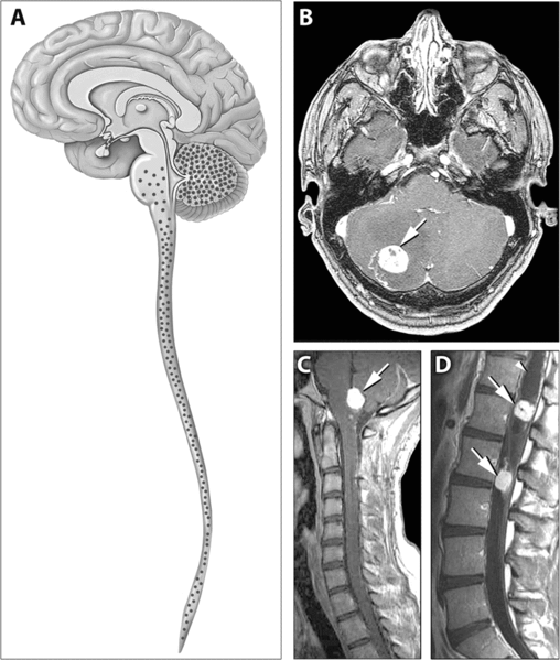
English: Distribution of Hemangioblastomas in the Central Nervous Systems of Study Patients

(A) Schematic representation of the distribution of CNS hemangioblastomas (red dots) in the 25 von Hippel-Lindau disease patients on MRI. Most (98%) of hemangioblastomas were found below the level of the tentorium in the cerebellum, brainstem, and spinal cord.

(B–D) Contrast-enhanced MRI demonstrating representative locations of hemangioblastomas including the cerebellum (B), brainstem (C) and spinal cord (D). (B) Axial view through the cerebellum demonstrating a hyperintense enhancing hemangioblastoma (arrow) with surrounding edema (hypointense area surrounding the tumor) that frequently is associated with these lesions. (C) Sagittal view through the posterior fossa demonstrating a hyperintense enhancing brainstem (medullary) hemangioblastoma (arrow) with surrounding edema. (D) Sagittal view through the thoracic and lumbar spinal cord demonstrating two hyperintense enhancing hemangioblastomas (arrows). The superior tumor is associated with a large intraspinal cyst (syrinx) that is common with these neoplasms (arrowhead)
Source http://medicine.plosjournals.org/perlserv/?request=get-document&doi=10.1371/journal.pmed.0040060
Auteur

Conditions d’utilisation

w:fr:Creative Commons
paternité
Ce fichier est disponible selon les termes de la licence Creative Commons Attribution 2.5 Générique.
Vous êtes libre :
  • de partager – de copier, distribuer et transmettre cette œuvre
  • d’adapter – de modifier cette œuvre
Sous les conditions suivantes :
  • paternité – Vous devez donner les informations appropriées concernant l'auteur, fournir un lien vers la licence et indiquer si des modifications ont été faites. Vous pouvez faire cela par tout moyen raisonnable, mais en aucune façon suggérant que l’auteur vous soutient ou approuve l’utilisation que vous en faites.
PLOS
PLOS
Cette image a été publiée dans un journal de la Public Library of Science. Leur site web indique que le contenu de tous les journaux de la PLOS sont publiés sous licence Creative Commons Attribution 2.5.

À celui qui importe l'image : Vous devez indiquer un lien (URL) vers l’image ou l’article d’origine.

Veuillez catégoriser l’image dans l’une des sous-catégories de Category:Media from PLOS journals.

Légendes

Ajoutez en une ligne la description de ce que représente ce fichier

Éléments décrits dans ce fichier

dépeint

image/gif

Historique du fichier

Cliquer sur une date et heure pour voir le fichier tel qu'il était à ce moment-là.

Date et heureVignetteDimensionsUtilisateurCommentaire
actuel31 mai 2007 à 15:43Vignette pour la version du 31 mai 2007 à 15:43805 × 949 (278 kio)Filip emDistribution of Hemangioblastomas in the Central Nervous Systems of Study Patients (A) Schematic representation of the distribution of CNS hemangioblastomas (red dots) in the 25 von Hippel-Lindau disease patients on MRI. Most (98%) of hemangioblastomas w

Aucune page n’utilise ce fichier.

Usage global du fichier

Les autres wikis suivants utilisent ce fichier :