
Fichier:Hepatocellular carcinoma 1.jpg
Hepatocellular_carcinoma_1.jpg (550 × 368 pixels, taille du fichier : 38 kio, type MIME : image/jpeg)
Ce fichier et sa description proviennent de Wikimedia Commons.
Description
| Description |
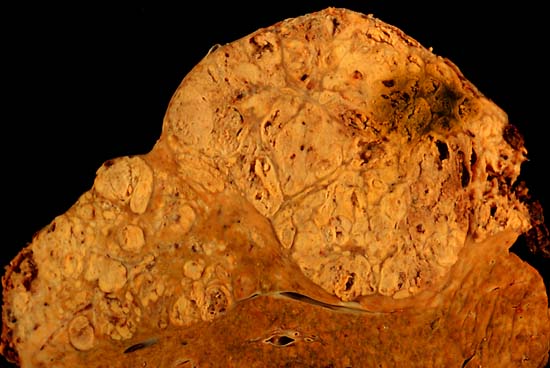
Hepatocellular carcinoma This specimen is from a 50ish woman who presented to the hospital with abdominal pain and ascites. The radiologist recovered what appeared to be whole blood on paracentesis. Cytological exam of the bloody fluid showed no evidence of malignancy. Liver function tests were abnormal, and serologic tests were positive for antibody to hepatitis C. The patient deteriorated rapidly and died within a few days. The autopsy showed this hepatocellular carcinoma occupying much of the volume of a cirrhotic liver. Furthermore, the tumor had invaded the diaphragm and ruptured into the peritoneal cavity, causing the bloody ascites. The photo shows a view of a longitudinal slice taken through the full length of the liver. The photos were shot with a Minolta X-370 with 100 mm bellows lens on Kodak Elite ISO 100 transparency film. The specimen was sliced fresh and fixed in formalin overnight, then briefly immersed in 70% alcohol to retrieve some of the native color and dull the surface reflections. Photograph by Ed Uthman, MD. Public domain. Posted 23 Sep 00 |
| Source | http://web2.airmail.net/uthman/specimens/index.html |
| Auteur | |
| Autorisation (Réutilisation de ce fichier) |
PD |
Conditions d’utilisation
| Cette œuvre a été placée dans le domaine public par son auteur, Ed Uthman. Ceci s’applique dans le monde entier. Dans certains pays, ceci peut ne pas être possible ; dans ce cas : Ed Uthman accorde à toute personne le droit d’utiliser cette œuvre dans n’importe quel but, sans aucune condition, sauf celles requises par la loi.
|
Historique du fichier
Cliquer sur une date et heure pour voir le fichier tel qu'il était à ce moment-là.
| Date et heure | Vignette | Dimensions | Utilisateur | Commentaire | |
|---|---|---|---|---|---|
| actuel | 5 juin 2006 à 12:14 | | 550 × 368 (38 kio) | Patho | {{Information| |Description=Hepatocellular carcinoma This specimen is from a 50ish woman who presented to the hospital with abdominal pain and ascites. The radiologist recovered what appeared to be whole blood on paracentesis. Cytological exam of the blo |
Utilisation du fichier
La page suivante utilise ce fichier :
Usage global du fichier
Les autres wikis suivants utilisent ce fichier :
- Utilisation sur ar.wikipedia.org
- Utilisation sur ast.wikipedia.org
- Utilisation sur az.wikipedia.org
- Utilisation sur be.wikipedia.org
- Utilisation sur bs.wikipedia.org
- Utilisation sur ca.wikipedia.org
- Utilisation sur cs.wikipedia.org
- Utilisation sur de.wikipedia.org
- Utilisation sur de.wikibooks.org
- Utilisation sur el.wikipedia.org
- Utilisation sur en.wikipedia.org
- Hepatocellular carcinoma
- Alcohol and cancer
- Portal:Medicine/Selected article/50, 2007
- Portal:Medicine/Selected Article Archive (2007)
- Obesity-associated morbidity
- Cirrhosis
- Portal:Viruses/Selected article
- Portal:Viruses/Selected article/10
- User:Daniel Mietchen/Wikidata lists/Items with Disease Ontology IDs
- Utilisation sur eo.wikipedia.org
- Utilisation sur es.wikipedia.org
- Utilisation sur eu.wikipedia.org
- Utilisation sur fa.wikipedia.org
- Utilisation sur fi.wikipedia.org
- Utilisation sur gl.wikipedia.org
- Utilisation sur he.wikipedia.org
- Utilisation sur hi.wikipedia.org
- Utilisation sur hy.wikipedia.org
- Utilisation sur id.wikipedia.org
- Utilisation sur it.wikipedia.org
- Utilisation sur ja.wikipedia.org
- Utilisation sur kk.wikipedia.org
Voir davantage sur l’utilisation globale de ce fichier.